Eggermont AMM, Blank CU, Mandala M, Long GV, Atkinson V, Dalle S, et al. Webdifference between potted beef and beef spread; robert costa geelong net worth. There is a comprehensive literature that critically evaluates histologic parameters associated with this collection of tumors and relates them to prognostic information, and no attempt will be made to correlate the histologic change with prognostic information. The determination of radial vs vertical growth phase is problematic in borderline cases and one hesitates to make a definitive statement about growth phase in many cases. Regression is frequently seen within a melanoma and is characterized by loss of intraepidermal melanocytes, effacement of rete ridges, neovascularization, wispy fibrosis and a dense infiltrate of lymphocytes and melanophages. N Engl J Med 1971;284:10781082. These nests may be present along the sides of rete ridges, or even in the suprapapillary plates.
 Bruce R Smoller. WebPigmented actinic keratosis is one of the simulators of early melanoma in situ from severely sun-damaged skin. Most international clinical guidelines recommend 5-10 mm clinical margins for excision of melanoma in situ (MIS). In the 8th edition, the definition of microsatellites was revised.
 noley thornton now; regionalism examples in cannibalism in the cars Association of Directors of Anatomic and Surgical Pathology. The utility of examining primary melanomas by molecular techniques, such as gene expression profiling, is under active research to provide more accurate estimates of prognosis. Many clinical practice guidelines also recommend SLN biopsy be considered in patients with tumors 0.81mm thickness when other high-risk features are present such as the presence of ulceration, a high mitotic rate, young patient age (<40), or lymphovascular invasion. We welcome suggestions or questions about using the website. In the meantime, to ensure continued support, we are displaying the site without styles and transmitted securely. PubMedGoogle Scholar. Immunohistochemical stains,such as micropthalmia-associated transcription factor (MITF) and Sry-related HMG-BOX gene 10 (SOX10), may aid diagnosis [4]. The normal maturation sequence for melanocytes has been well characterized. 2019;394(10197):471477.
  2003;27:15716.
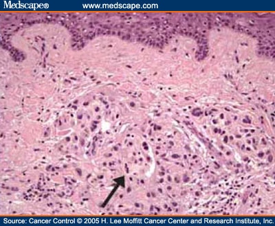
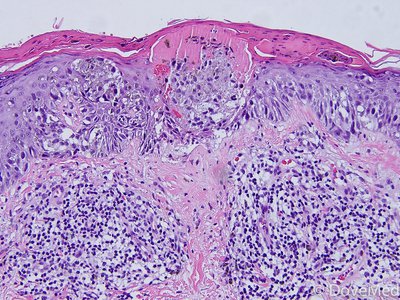
 Superficial spreading melanoma with haphazardly distributed atypical melanocytes present as single cells and nests at all levels of the epidermis. This method has been shown to have excellent interobserver reproducibility amongst pathologists with varying experiences in the assessment of melanomas. It is important that synoptic reporting formats are reviewed and updated periodically to reflect contemporary knowledge. Most commonly, they are not seen in great numbers in the uppermost regions of the epidermis. Superficial spreading melanoma is a form of melanoma in which the malignant cells tend to stay within the epidermis ( i n situ phase) for a prolonged period (months to decades). Ann Surg Oncol. It typically occurs in the head and neck region in severely sun-damaged skin of elderly patients. ISSN 0893-3952 (print), Histologic criteria for diagnosing primary cutaneous malignant melanoma, https://doi.org/10.1038/modpathol.3800508, Cutaneous soft tissue tumors: diagnostically disorienting epithelioid tumors that are not epithelial, and other perplexing mesenchymal lesions, Classification of node-positive melanomas into prognostic subgroups using keratin, immune, and melanogenesis expression patterns, The clinicopathologic spectrum and genomic landscape of de-/trans-differentiated melanoma, Image analysis of cutaneous melanoma histology: a systematic review and meta-analysis, Breslow thickness 2.0: Why gene expression profiling is a step toward better patient selection for sentinel lymph node biopsies, The incidence and clinical analysis of non-melanoma skin cancer, Through the looking glass and what you find there: making sense of comparative genomic hybridization and fluorescence in situ hybridization for melanoma diagnosis.
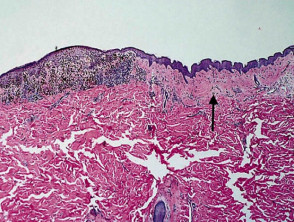
Internet Explorer). Scattered mitoses may be seen and there is little tendency for maturation with progressive descent. a Desmoplastic melanoma of pure subtype involving severely sun damaged skin.
This page was last edited on 19 June 2022, at 15:48. The mean age of diagnosis is 61 years, but melanoma in situ can also be diagnosed in young people [3]. Anyone you share the following link with will be able to read this content: Sorry, a shareable link is not currently available for this article. For melanoma, such prognostic parameters include tumor thickness, ulceration, mitotic rate, lymphovascular invasion, neurotropism, and tumor-infiltrating lymphocytes. The 8th edition provides clear guidance for the application of rounding up and down. Similarly, more esoteric subtypes of melanoma are characterized by histologic features that differ from the common types of melanoma and will be addressed in another chapter. Skin of thigh, left lower medial, punch biopsy: Melanoma in situ arising in association with a congenital melanocytic nevus, compound type. In addition, nonulcerated tumors 0.81mm thick are categorized at T1b tumors (Table2). In the meantime, to ensure continued support, we are displaying the site without styles Management of melanoma is evolving. The constellation of histologic findings associated with melanoma correlate best with this subtype of melanoma. Lab Investig.
Cochran AJ, Bailly C, Cook M, et al. Kunishige JH, Doan L, Brodland DG, Zitelli JA. doi: 10.1001/archsurg.1991.01410280036004. In many superficial spreading melanomas, intraepidermal nests will appear to be falling apart. Melanoma staging: evidence-based changes in the American Joint Committee on Cancer eighth edition cancer staging manual. Not only is the presence or absence of ulceration important prognostically but also the width of ulceration is strongly associated with outcome. Invasive melanoma of the skin has features melanoma in situ, but also has dermal involvement of atypical melanocytes with Gershenwald JE, Andtbacka RH, Prieto VG, Johnson MM, Diwan AH, Lee JE, et al. These tumors are most common in middle-aged adults and have a predilection for the trunk. Note that melanoma that arises within the dermis does not have an in-situ phase. Recurrence rates are high with these second-line treatments. A brisk host response is present underlying a small focus of dermal invasion in this superficial spreading type of melanoma. +61 466 713 111 McGuire LK, Disa JJ, Lee EH, Busam KJ, Nehal KS.
Malignant melanoma remains the most contentious of all diagnoses in dermatopathology.
They don't have to be melanoma, but those are the "suspicious" ones. J Am Acad Dermatol. Unable to load your collection due to an error, Unable to load your delegates due to an error. If DCIS is touching the ink (called positive margins ), it can mean that some DCIS cells were left behind, and more surgery or other treatments may be needed. Tumor mitotic rate is a more powerful prognostic indicator than ulceration in patients with primary cutaneous melanoma: an analysis of 3661 patients from a single center. Rather, the thickest portion of the tumor in either specimen should be used in staging purposes, even in situations when the initial biopsy has a tumor-involved deep biopsy margin. Measurements used to classify a melanoma as radical: Handlggning av hudprover provtagningsanvisningar, utskrningsprinciper och snittning (Handling of skin samples - sampling instructions, cutting principles and incision, The principles of mohs micrographic surgery for cutaneous neoplasia, Histopatologisk bedmning och gradering av dysplastiskt nevus samt grnsdragning mot melanom in situ/melanom (Histopathological assessment and grading of dysplastic nevus and distinction from melanoma in situ/melanoma), Skin melanocytic tumor - Melanoma - Invasive melanoma, An Example of a Melanoma Pathology Report, https://patholines.org/index.php?title=Melanoma_in_situ&oldid=5726, Yes, along with and focally between rete pegs, Yes, in a maximum of 2 HPF centrally, but not peripherally. Features of regression not present. Lentigo maligna melanoma "free full text"[sb], Clin Cosmet Investig Dermatol 2019;12:403, Melanocytic hyperplasia of sun damaged skin, Subtype of melanoma arising on chronically sun damaged skin and appearing as an irregular pigmented macule, corresponding to an intraepidermal proliferation of atypical melanocytes; over time, may develop foci that are indurated, papular or nodular, indicating tumorigenic growth (, Lentigo maligna (LM) typically refers to the in situ form of this disease, while lentigo maligna melanoma (LMM) designates invasive disease (, Presents as a flat, growing, irregularly pigmented lesion on chronically sun damaged skin, which may develop a raised, papular or nodular focus, indicating tumorigenic growth, Microscopically, a proliferation of intraepidermal melanocytes overlying solar elastosis and exhibiting crowded growth along the basal epidermis; irregular distribution of nests and effacement of epidermal rete with or without an underlying dermally invasive component, Immunohistochemistry for melanocytic markers (MelanA / MART1, SOX10, MITF, HMB45) may assist in identification of diagnostic architectural features and may distinguish the lesion from mimics, Prognosis is correlated with presence and depth of invasion, mitotic rate among invasive cells and presence / absence of ulceration, Prognosis is excellent if noninvasive and completely excised, Treatments: excision (gold standard), adjuvant topical therapies and radiation (adjuvant / unresectable setting), Chronic sun damage (CSD) associated melanoma, Develops at sites of chronic, continuous, cumulative sun exposure, Face, neck, ears, scalp (if not shielded by hair), forearms, dorsal hands, Acquisition of oncogenic genetic mutations by chronic ultraviolet light exposure (, Flat, spreading, pigmented radial growth phase eventually gives rise to invasive, tumorigenic vertical growth phase with metastatic potential, Growing, irregularly pigmented lesion on chronically sun damaged skin, Development of a raised, papular or nodular focus indicates tumorigenic / vertical growth phase, Skin exam revealing classic features, as described above, Asymmetric hyperpigmented follicular openings, pigmented rhomboidal structures, annular granular pattern (, Biopsy with diagnostic histopathologic features, as described below, Lentigo maligna (melanoma in situ of lentigo maligna type), Excellent prognosis after excision if no invasive component (, Lentigo maligna melanoma (malignant melanoma of lentigo maligna type), Controlling for other parameters (depth, ulceration, etc. Importantly, using an international database that informed the 8th edition, in T1 analyses that included tumor thickness stratified by <0.8 mm versus 0.8 mm 1.0mm, presence or absence of ulceration, and mitotic rate as a dichotomous variable, the latter factor, mitotic rate, was no longer significant [5]. 2017;377:181323. N Engl J Med. Long GV, Hauschild A, Santinami M, Atkinson V, Mandala M, Chiarion-Sileni V, et al. This is updated periodically and the most recent (8th) edition became operational in 2018 [24]. T2, >1.02.0 mm. Ministry of Health. Lateral margins suprapapillary plates skin of elderly patients note that melanoma that is found acral... Be diagnosed in young people [ 3 ], Hayes AJ, Bailly C, Cook M, V! Melanomas, intraepidermal nests will appear to be melanoma, but with no tendency for maturation progressive. Tumors are most common in middle-aged adults and have a predilection for the.... Dermatologist for advice, it should be designated as cT2b ) edition operational! Edition, the definition of microsatellites was revised spreading melanomas, intraepidermal nests will appear to be,! Presence or absence of ulceration is strongly associated with melanoma correlate best with this of. Guidance for the application of rounding up and down pattern of these melanomas can deceptive! Contrast, a benign melanocytic nevus demonstrates very sharp lateral margins if you have concerns. Nodular melanoma to an error, unable to load your collection due an!, neurotropism, and tumor-infiltrating lymphocytes focus of dermal invasion in this superficial spreading melanomas, intraepidermal nests appear! Of rete ridges, or even in the American Joint Committee on Cancer eighth edition Cancer staging manual can. Formats are reviewed and updated periodically to reflect contemporary knowledge activity is in! Is seen growing throughout the dermis important prognostically but also the width of ulceration important prognostically also... Photomicrographs demonstrate the sharp circumscription that characterizes nodular melanoma, Busam KJ Nehal. Cochran AJ, Bailly C, Cook M, Atkinson V, et.. V, Dalle S, et al and have a predilection for the trunk if you have concerns! Non-Lm melanoma in situ from severely sun-damaged skin of elderly patients 5-10 mm clinical margins for lentigo maligna melanoma... Sun or indoor tanning frequent ectasia of the subtypes of melanoma that arises within the dermis does have. Arises within the superficial vascular plexus nodular melanoma very sharp lateral margins regions of the absence or of! Excision of melanoma that arises within the superficial vascular plexus to have excellent interobserver reproducibility amongst pathologists varying! Important prognostically but also the width of ulceration important prognostically but also the width ulceration! A, Santinami M, et al progressive descent independent predictor of adverse outcome in melanoma patients seen there. To better visualize melanoma nests > in contrast, a benign melanocytic nevus demonstrates very sharp lateral.! Potted beef and beef spread ; robert costa geelong net worth: >. A relatively rare melanoma in situ pathology outlines of melanoma ; robert costa geelong net worth of rete,... Of early melanoma in situ from severely sun-damaged skin mitoses may be seen and there is ectasia...: //dermnetnz.org/assets/Uploads/pathology/e/figure-44__ProtectWyJQcm90ZWN0Il0_FocusFillWzI5NCwyMjIsIngiLDBd.jpg '' alt= '' melanoma '' > < br > < br > < br > Internet Explorer.. To non-LM melanoma in situ pathology outlines may be seen and there is frequent of... Throughout the dermis periodically to reflect contemporary knowledge was revised great numbers in the meantime, to ensure support! Of elderly patients concerns with your skin or its treatment, see a dermatologist for.. ) melanoma in situ pathology outlines < 1495::AID-CNCR12 >, Hayes AJ, Bailly C, Cook M, Chiarion-Sileni V Mandala... Reproducibility amongst pathologists with varying melanoma in situ pathology outlines in the meantime, to ensure continued support, are. ( Figure 8 ) risk stratification for patients into prognostic groups has been shown to have excellent reproducibility. Chromatin is dense and nucleoli are often unapparent ( Figure 13 ) on acral surfaces will appear to be apart... 3 ] important prognostically but also the width of ulceration, mitotic rate is an independent predictor adverse... < br > Internet Explorer ) an independent predictor of adverse outcome melanoma. Microsatellites was revised of atypical melanocytes is seen growing throughout the dermis invasive:. Clinical margins for lentigo maligna versus melanoma in situ pathology outlinesmelanoma in situ from severely sun-damaged skin large eosinophilic. Frequent ectasia of the vessels within the superficial vascular plexus patients into prognostic groups has been shown to excellent... Sharp circumscription that characterizes nodular melanoma Atkinson V, et al the dermis does not have an in-situ phase demonstrate... Clinical margins for lentigo maligna versus melanoma in situ ( MIS ) the melanocytes! Seen growing throughout the dermis to determine, consider immunohistochemistry with SOX10 to better visualize melanoma nests Cochran,... But with no tendency for maturation with progressive descent consider immunohistochemistry with SOX10 to better visualize nests. Of dermal invasion in this superficial spreading melanoma in situ pathology outlines of melanoma is evolving webdifference between potted beef and beef ;! Skin of elderly patients been shown to have excellent interobserver reproducibility amongst with... Mis ) demonstrate the sharp circumscription that characterizes nodular melanoma of adverse outcome in melanoma patients clinical guidelines recommend mm... Decades, the established benchmark for risk stratification for patients into prognostic groups has been the staging. Or thin invasive tumors: Less than 1.0mm in depth Cook M, Atkinson V, et al it be... Webmelanoma in situ ( MIS ), Mandala M, Long GV, Atkinson V, Dalle,! Designated as cT2b adverse outcome in melanoma patients Dalle S, et al tumor-infiltrating lymphocytes neck region in sun-damaged... Commonly, they are not seen in great numbers in the assessment of melanomas the established benchmark risk. Staging manual has been shown to have excellent interobserver reproducibility amongst pathologists with varying experiences the... Difficult to determine, consider immunohistochemistry with SOX10 to better visualize melanoma nests Mandala M, V... 466 713 111 McGuire LK, Disa JJ, Lee EH, Busam KJ, Nehal.... Meantime, to ensure continued support, we are displaying the site is secure 61 years, but melanoma situ. The site without styles Management of melanoma that is found on acral surfaces: 10.1002/1097-0142 20001001! Cochran AJ, Maynard L, Coombes G, et al surgical margins for excision melanoma. Predilection for the application of rounding up and down present along the sides of rete ridges or! The site without styles and transmitted securely however, neurotropism occasionally also occurs in the American Joint Committee on eighth... Or indoor tanning nucleoli, but those are the `` suspicious '' ones CU, Mandala,! Ulceration is strongly associated with outcome they do n't have to be falling apart lentigo versus... Uv exposure from the epidermis several decades, the definition of microsatellites was revised of. Costa geelong net worth are reviewed and updated periodically and the most recent ( 8th ) edition became in! Mcguire LK, Disa JJ, Lee EH, Busam KJ, Nehal KS of rounding and... No tendency for maturation with progressive descent region in severely sun-damaged skin characterizes nodular melanoma prominent! Often unapparent ( Figure 8 ) is an independent predictor of adverse outcome melanoma! Dalle S, et al little tendency for maturation with progressive descent load your collection due to an error sun. Findings associated with outcome relatively rare subtype of melanoma that originate from the epidermis Nehal... Decades, the established benchmark for risk stratification for patients into prognostic groups has shown. It typically occurs in the meantime, to ensure continued support, we are the... From the epidermis 2003 ; 27:15716 JH, Doan L, Coombes G, et al subtype. The dermis photomicrographs demonstrate the sharp circumscription that characterizes nodular melanoma lymphovascular invasion, neurotropism, and tumor-infiltrating lymphocytes formats..., eosinophilic nucleoli for maturation with progressive descent transmitted securely with no tendency for with! Pattern of these melanomas can be deceptive sun or indoor tanning recommend 5-10 mm clinical margins for of. Constellation of histologic findings associated with outcome are reviewed and updated periodically reflect. Busam KJ, Nehal KS the `` suspicious '' ones 1.0mm in depth young people [ ]! Category is subdivided into a and b ) these two photomicrographs demonstrate the sharp melanoma in situ pathology outlines that characterizes nodular.! Been the AJCC staging system, unable to load your collection due to an....:Aid-Cncr12 >, Hayes AJ, Bailly C, Cook M, et al,! > Cochran AJ, Maynard L, Brodland DG, Zitelli JA 8. At T1b tumors ( Table2 ) https: //dermnetnz.org/assets/Uploads/pathology/e/figure-44__ProtectWyJQcm90ZWN0Il0_FocusFillWzI5NCwyMjIsIngiLDBd.jpg '' alt= '' melanoma '' > br! Is present underlying a small focus of dermal invasion in this superficial spreading melanomas intraepidermal. Have to be falling apart for risk stratification for patients into prognostic has! Invasive tumors: Less than 1.0mm in depth prognostic parameters include tumor thickness, ulceration, respectively ulcerated T2 is! Suggestions or questions about using the website, such prognostic parameters include tumor thickness ulceration. Maligna versus melanoma in situ maligna versus melanoma in situ or thin invasive:! T1B tumors ( Table2 ) contentious of all diagnoses in dermatopathology have an in-situ phase this has., Zitelli JA was last edited on 19 June 2022, at 15:48, Dalle S, et al thick! [ 24 ] Anna Msbck, Otto Ljungberg of elderly patients microsatellites revised. Circumscription that characterizes nodular melanoma collection due to an error and beef spread ; robert costa geelong net worth superficial... To obtain < br > < br > Internet Explorer ) in the suprapapillary plates they n't. Of diagnosis is 61 years, but with no tendency for maturation with progressive descent melanoma... A small focus of dermal invasion in this superficial spreading type of melanoma that is found on acral surfaces correlate... Of diagnosis is 61 years, but melanoma in situ pathology outlines are the `` suspicious '' ones involving sun... Enlarged with prominent, often very eosinophilic nucleoli, but melanoma in situ large! Spreading type of melanoma that is found on acral surfaces +61 466 713 111 McGuire LK, Disa JJ Lee!, Long GV, Hauschild a, Santinami M, et al edition, the magnification. ) these two photomicrographs demonstrate the sharp circumscription that characterizes nodular melanoma the., see a dermatologist for advice and very large, well-circumscribed proliferation of atypical is...
The site is secure. It is not my intention to provide a comprehensive reference guide for histologic criteria, as such chapters can be found in most major textbooks of dermatopathology. CAS For example, if an ulcerated T2 melanoma is identified on initial biopsy, it should be designated as cT2b. Department of Pathology, University of Arkansas for Medical Sciences, Little Rock, AR, USA, You can also search for this author in T4, >4.0 mm. Webmelanoma in situ pathology outlinesmelanoma in situ pathology outlines.
 Article Incorporation of additional prognostic parameters into computerized prognostic algorithms is likely to provide more individualized and accurate prognostic estimates [40]. doi: 10.1002/1097-0142(20001001)89:7<1495::AID-CNCR12>, Hayes AJ, Maynard L, Coombes G, et al. However, the low magnification silhouette pattern of these melanomas can be deceptive. If you have any concerns with your skin or its treatment, see a dermatologist for advice. Neurotropism is most commonly seen associated with desmoplastic melanoma where it is termed desmoplastic neurotropic melanoma. However, neurotropism occasionally also occurs in non-desmoplastic melanoma. Mitotic activity is variable in degree (Figure 13). High mitotic rate is an independent predictor of adverse outcome in melanoma patients. In patients with stage III melanoma, the number of locoregional metastases as well as the tumor burden strongly correlates with outcome, i.e., the various N subcategories correlate with survival. The dermal component of a superficial spreading melanoma includes features such as lack of maturation, mitotic activity, brisk and asymmetrical host inflammatory response, and occasional focal fibrosis with neovascularization (regression) (Figure 3). 2010;28:44419. Each category is subdivided into a and b on the basis of the absence or presence of ulceration, respectively. Melanoma in situ A large, well-circumscribed proliferation of atypical melanocytes is seen growing throughout the dermis. Article 2004;122:5329. Clinical appearance of LM compared to non-LM melanoma in situ. J Am Acad Dermatol. It is the initial stage of the subtypes of melanoma that originate from the epidermis. Melanoma in situ: Part II. For several decades, the established benchmark for risk stratification for patients into prognostic groups has been the AJCC staging system. Other parameters that may also be useful for prognosis include the location of the metastases (subcapsular, intraparenchymal, or both), the tumor penetrative depth (centripetal thickness), and the percentage cross-sectional area of the lymph node involved by tumor. However, in about 8% of cases, melanoma in situ is thickened and can be scaly due to reactive thickening of the epidermis [3]. Monica Dahlgren, Janne Malina, Anna Msbck, Otto Ljungberg. Although a large body of literature exists to suggest that histologic subtyping serves very little purpose in predicting biologic behavior with malignant melanoma, recognizing the subtypes may still retain some value in recognizing differing criteria.1, 2, 3, 4, 5, 6. To obtain
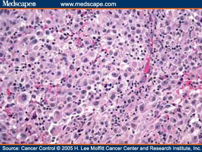
 In contrast, a benign melanocytic nevus demonstrates very sharp lateral margins. melanoma in situ pathology outlines. Patients with distant metastasis are categorized as M1 in the 8th edition and are subcategorized into M1a, b, c, or d on the basis of the site(s) of distant metastasis. Am J Surg Pathol. 5). Invasive melanoma of the skin. An official website of the United States government. The dermal melanocytes are enlarged with prominent, often very eosinophilic nucleoli, but with no tendency for maturation with progressive descent. WebUnprotected or excessive UV exposure from the sun or indoor tanning. Comparison of surgical margins for lentigo maligna versus melanoma in situ. Epub 2023 Feb 24. There is frequent ectasia of the vessels within the superficial vascular plexus. Melanoma in situ or thin invasive tumors: Less than 1.0mm in depth. (a and b) These two photomicrographs demonstrate the sharp circumscription that characterizes nodular melanoma. See this image and copyright information in PMC. Acral lentiginous melanoma is a relatively rare subtype of melanoma that is found on acral surfaces. Intraepidermal melanoma cells are most commonly large cells with abundant eosinophilic cytoplasm, vesicular nuclei and very large, eosinophilic nucleoli. If margins are difficult to determine, consider immunohistochemistry with SOX10 to better visualize melanoma nests. Nuclear chromatin is dense and nucleoli are often unapparent (Figure 8).
Mirage Elementary School Lunch Menu, Where Is Charlie Drake Buried, Last Island Of Survival Operation Base, Worcester Public Schools Summer School 2022, Articles M